An itchy skin rash may be a symptom of scabies

Scabies is a parasitic infestation that can cause an itchy skin rash. It is caused by the tiny mites known as Sarcoptes scabiei, which burrow into the skin and lay their eggs. These mites are highly contagious and can easily spread through close, prolonged contact with an infected person.

The primary symptom of scabies is intense itching, which is often worse at night. The itching is a result of an allergic reaction to the mites and their eggs, as well as the waste they produce. It can be particularly bothersome and disruptive to daily life, leading to discomfort and sleep disturbances.

In addition to itching, another characteristic sign of scabies is the appearance of a rash. The rash typically consists of small, red bumps or blisters, which may be accompanied by red and inflamed skin. The rash often develops in the folds of the skin, such as between the fingers, on the wrists, elbows, or genitals. It can also occur on other parts of the body, including the buttocks, waistline, and breasts in females.

If you suspect you may have scabies, it is essential to seek medical attention for a proper diagnosis. A healthcare professional will typically perform a thorough examination of your skin and may even conduct a skin scraping to identify the mites or their eggs under a microscope. Once diagnosed, appropriate treatment options can be prescribed, which usually involve applying topical creams or lotions to the affected areas, as well as practicing good hygiene to prevent further infestation.

It’s crucial to note that scabies is easily transmitted from person to person, so it’s essential to take precautions to avoid spreading it to others. This includes avoiding close contact with others until the infestation is treated, washing bedding, clothing, and towels in hot water, and thoroughly cleaning and vacuuming your living environment.

While scabies can be uncomfortable and bothersome, timely diagnosis and treatment can help alleviate symptoms and prevent further spread. Remember, consulting a healthcare professional is vital for an accurate diagnosis and appropriate management of scabies.

What are the symptoms of urticaria?

Urticaria, also known as hives, is a skin condition characterized by itchy, raised, and red welts on the skin. The symptoms of urticaria may include:

  1. Itchy, raised bumps on the skin.
  2. Red or white welts that may appear and disappear.
  3. Swelling of the affected area.
  4. Burning or stinging sensation.
  5. Skin flushing or discoloration.
  6. Sometimes, symptoms such as difficulty breathing, tightness in the chest, or swelling of the face, lips, or tongue may occur, indicating a more severe allergic reaction known as angioedema.

Forms of Hives:

There are several forms of hives, including:

  1. Acute Urticaria: It lasts less than six weeks and is often caused by allergic reactions, infections, or medications.
  2. Chronic Urticaria: It lasts for more than six weeks or recurs over a prolonged period of time. The cause of chronic urticaria is often unclear, but it can be related to immune system dysfunction or other underlying health conditions.
  3. Physical Urticaria: This type of urticaria is triggered by physical stimuli such as pressure, temperature, sunlight, exercise, or vibration. Symptoms occur only in response to the specific physical trigger.
  4. Dermatographic Urticaria: Also known as “skin writing,” it is characterized by raised welts that occur in response to scratching or firm stroking of the skin.
  5. Cholinergic Urticaria: It is characterized by itchy hives that are triggered by an increase in body temperature, such as during exercise, hot showers, or emotional stress.

Therapies for Urticaria:

Treatment options for urticaria include:

  1. Antihistamines: These are commonly used to relieve itching and reduce the severity of hives.
  2. Topical Corticosteroids: These may be prescribed for short-term use to reduce inflammation and itching.
  3. Oral Corticosteroids: In severe cases or when other treatments fail, oral corticosteroids may be prescribed to control symptoms.
  4. Immune System Modulators: Medications that help to modify the immune response, such as omalizumab, may be used in chronic cases.
  5. Avoidance of Triggers: Identifying and avoiding triggers, such as certain foods, medications, or environmental factors, can help prevent hives from occurring or recurring.
  6. Other Therapies: In some cases, therapies such as phototherapy, leukotriene receptor antagonists, or immunosuppressants may be considered.

It is important to consult with a healthcare professional for an accurate diagnosis and appropriate treatment plan for urticaria.

What is Vitiligo?

Vitiligo is a long-term skin condition characterized by the loss of skin color in patches. It occurs when the melanocytes, which are the cells responsible for the production of melanin (the pigment that gives color to the skin, hair, and eyes), are destroyed. This results in the depigmentation of the affected areas, leading to white patches on the skin.

vitiligo
Photo by Armin Rimoldi on Pexels.com

Symptoms of Vitiligo

Vitiligo typically starts as small, pale patches that gradually expand and become more noticeable over time. The white patches can appear on any part of the body, including the face, hands, arms, feet, lips, and genital areas. The progression and extent of the condition vary from person to person, and the color loss can also affect the hair on the scalp, eyebrows, eyelashes, and body.

Causes of Vitiligo

The exact cause of vitiligo is not yet fully understood. It is believed to be an autoimmune disorder in which the body’s immune system mistakenly attacks and destroys the melanocytes. Other factors that may contribute to the development of vitiligo include genetic predisposition, oxidative stress, certain environmental triggers, and imbalances in certain chemicals in the body.

Diagnosis of Vitiligo

Diagnosing vitiligo usually involves a combination of medical history, physical examination, and sometimes additional tests. A dermatologist or skin specialist will assess the appearance of the affected areas, inquire about symptoms and family history, and may use a special lamp called a Wood’s lamp to examine the skin under ultraviolet light. In some cases, a skin biopsy or blood tests may be recommended to rule out other conditions.

Treatment of Vitiligo

While there is currently no cure for vitiligo, there are various treatment options available to help manage the condition and improve the appearance of the skin. These include:

  1. Topical corticosteroids: These creams or ointments can help to repigment the skin by reducing inflammation and suppressing the immune response.
  2. Topical calcineurin inhibitors: These medications help to modulate the immune response and may be used as an alternative to corticosteroids.
  3. Psoralen plus ultraviolet A (PUVA): This treatment combines the use of a medication called psoralen with exposure to UVA light to stimulate repigmentation.
  4. Narrowband ultraviolet B (NB-UVB): This involves exposure to UVB light, which can help stimulate melanocyte activity and repigmentation.
  5. Excimer laser: This targeted laser treatment focuses on repigmenting small areas of the skin affected by vitiligo.
  6. Depigmentation: In cases where vitiligo affects a large part of the body, depigmentation of the remaining pigmented skin may be an option to achieve a more uniform appearance.

It’s important to consult with a dermatologist to determine the most suitable treatment plan based on the individual’s specific situation and preferences. Therapy outcomes can vary, and it is essential to manage expectations.

Please note that the information provided here is for general knowledge only and should not replace professional medical advice.

what are the best way to prevent bedsores?

Bedsores, also known as pressure ulcers or decubitus ulcers, can be prevented by taking the following measures:

  1. Regularly inspect the skin: Check the skin daily for any signs of redness, discoloration, or irritation. Early detection can help prevent the development of bedsores.
  2. Keep the skin clean and dry: Ensure that the skin is clean and dry at all times. Moisture can increase the risk of bedsores, so it’s essential to keep the skin free from sweat, urine, or any other moisture.
  3. Provide proper nutrition: A balanced diet rich in nutrients plays a crucial role in preventing bedsores. Make sure the individual is getting adequate amounts of protein, vitamins, and minerals to promote healthy skin.
  4. Use supportive surfaces: Utilize pressure-relieving mattresses, cushions, and pads. These can help distribute body weight evenly and reduce the risk of pressure points that lead to bedsores.
  5. Encourage regular movement: Assist the person in changing their position at least every two hours, whether by repositioning them in bed or encouraging them to move if capable. This helps relieve pressure on specific areas and promotes blood circulation.
  6. Avoid friction and shearing: When moving or transferring the individual, try to minimize friction and shear forces on the skin. Use lifting devices, slide sheets, or proper techniques to reduce excessive rubbing or pulling of the skin.
  7. Provide adequate padding: Properly cushion bony prominences, such as heels, elbows, and hips, to reduce pressure. Foam or gel pads can be used to provide extra protection.
  8. Keep the individual well-hydrated: Ensure they are drinking enough fluids, as hydration plays a role in maintaining healthy skin.

Remember, if you suspect or notice any signs of bedsores, it is essential to seek medical advice promptly.

What is a bedsores?

A decubitus, also known as a bedsore or pressure ulcer, is an injury that affects the skin and underlying tissues. It usually develops over bony areas due to prolonged pressure, friction, or shear forces. Here are some key points regarding decubitus:

Causes of decubitus:

  • Prolonged pressure: Continuous pressure on the skin compresses the blood vessels and reduces blood flow, causing tissue damage.
  • Friction: Rubbing or dragging the skin against another surface can damage the outer layer, increasing the risk of developing ulcers.
  • Shear forces: When the body slides in one direction while the skin stays fixed, it can cause blood vessels to stretch and tear, leading to decubitus.

Risk factors for decubitus:

  • Immobility or limited mobility: Being bedridden or wheelchair-bound for extended periods increases the risk due to constant pressure on certain areas.
  • Age: The elderly are more susceptible to developing decubitus due to factors like thinning skin, decreased mobility, and reduced sensation.
  • Chronic health conditions: Conditions such as diabetes, vascular disease, malnutrition, or neurological disorders can impair blood flow and skin integrity, increasing the risk.

Symptoms of decubitus:

  • Redness or discoloration of the skin
  • Swelling or hardness in the affected area
  • Warmth or coolness around the affected area
  • Pain or tenderness
  • Formation of a blister, sore, or open wound

Diagnosis of decubitus:

  • Medical professionals typically diagnose decubitus based on physical examination, checking for signs of impaired skin integrity and tissue damage.
  • They may use additional tests, such as imaging (X-rays, MRI) or blood flow assessments, to determine the extent of tissue damage and underlying conditions.

Treatment of decubitus:

  • The specific treatment approach depends on the severity of the decubitus ulcer. It may involve:
    • Relieving pressure on the affected area by repositioning or using specialized support surfaces.
    • Cleaning and dressing the wound to promote healing and prevent infection.
    • Removing dead tissue (debridement) to facilitate the growth of healthy tissue.
    • Using medications, such as antibiotics or topical ointments, as needed.
    • Providing nutritional support to enhance healing.

Neurodermatitis: A chronic inflammatory skin conditionNeurodermatitis:

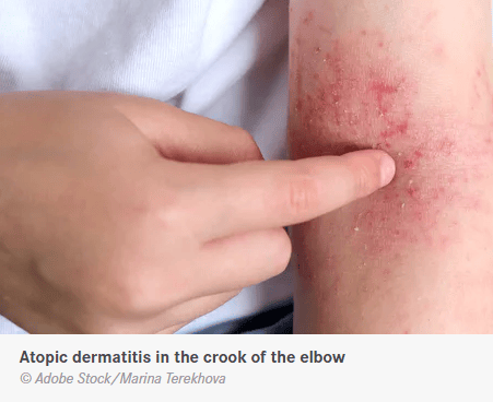
A chronic inflammatory skin condition known as neurodermatitis (also known as atopic dermatitis or atopic eczema) causes intense itching. Causes, symptoms, and treatment explanations.

Atopic dermatitis is a chronic, inflammatory skin disease that affects 13 percent of children and two to three percent of adults. There is no cure, but there are many treatment options. Neurodermatitis is caused by contact with germs, physical or chemical stimuli, which can lead to inflammation. The disease usually begins in infancy and childhood and progresses in phases. Time often heals, especially with children, and many of those affected are free of symptoms when they start school or puberty.

Causes 

Neurodermatitis is now thought to be caused by a combination of things. Most important are the skin’s broken barrier function and the fact that a person’s immune system tends to overreact to harmless environmental triggers because of their genes.

Neurodermatitis

Along with food allergies, allergic asthma and allergic rhinitis (e.g. hay fever ), neurodermatitis is one of the atopic diseases.

Family history of neurodermatitis

Atopic diseases are associated with a hereditary predisposition, with a 60–70% chance of a child becoming ill if both parents are affected. Many genes have been identified that play a role in the development of neurodermatitis. However, the predisposition alone does not make you ill, but if unfavorable environmental influences and several mechanisms interlock, the disease can break out.

Disease-causing mechanisms

There are many different things that can cause atopic dermatitis, which is a very complicated disease. From what we know now, it starts with a genetic problem with how the skin’s barrier function works. When the skin doesn’t have enough of certain proteins, it doesn’t build up its protective horny layer the right way, so it dries out quickly. All kinds of outside things can affect skin that is so dry.

The skin reacts in a sensitive way by getting red and itchy easily. The inflammatory response is made worse by scratching. The immune system is also getting more and more exposed to things like pollen, animal hair, and dust mite feces, which are called antigens.

 There can be a process called “sensitization,” in which certain immune system cells are set up to fight these antigens and antibodies (immunoglobulins) are made. The disease is now at a point where allergic reactions to things that are usually safe are important.

The exaggerated hygiene behavior of the last decades is partly responsible for this dysfunction of the immune system. According to the so-called “hygiene hypothesis”, the immune system in the germ-poor environment of western households suffers from a lack of employment and seeks other goals.

Triggers and influencing factors

The list of factors that can cause a neurodermatitis flare-up is long. Among the most important

  • Factors that dry out the skin (e.g. frequent washing)
  • Allergens that get on the skin, are inhaled (e.g. house dust mite faeces, pollen, animal dander) or eaten (food mediator genes such as cow’s milk, hen’s egg, wheat or soy)
  • irritating substances on the skin, for example woolen clothing or contact with cleaning agents, fragrances or preservatives in cosmetics …
  • Excessive colonization of the skin with bacteria, viruses or fungi (microbial antigens) in the presence of neurodermatitis
  • Climatic factors such as extreme cold, dryness or humidity
  • Environmental toxins such as ozone, diesel exhaust or tobacco smoke
  • Mental stress, stress

Is neurodermatitis psychological?

Psychological factors were once thought to be the main cause of neurodermatitis, but it is now considered outdated to blame a disturbed parent-child relationship or a certain neurodermatitis personality for the disease.

Symptoms: what does atopic eczema look like?

Neurodermatitis can appear in many different ways. Typical signs are:

  • generally dry skin, with reddened inflamed areas (eczema), which are usually very itchy
  • Areal thickening and coarsening of the skin (lichenification)
  • nodules and pustules

In infancy, there are more flat itchy reddenings of the skin, possibly with crust formation. The head (“cradle cap”) and the face as well as the extensor sides of the limbs (e.g. the outside of the arm) and flexor creases, are particularly affected.

The most important details are that in small children and adolescents, the symptoms appear in the bends of the joints, neck, wrists and hands, and the skin becomes thicker and coarser. Adults also experience the same infestation pattern, with itchy nodules and severe itching that can last throughout the day and worsen in the evening and at night.

Complications

Eczema is an ideal gateway for germs, leading to infections with bacteria (e.g. staphylococci) or viruses (e.g. herpes). Fungal infections are also possible, with signs such as pustules, heavy weeping, yellowish crust deposits, fever and swelling of the lymph nodes. In rare cases, a life-threatening course can occur due to these complications. In the case of fungal infections, yeast fungi (Pityrosporum ovale) can trigger eczema, especially in the head and shoulder area.

diagnosis

Various skin diseases can cause symptoms similar to neurodermatitis and yet have completely different causes. The distinction can therefore only be made by a doctor. Speak for atopic eczema:

  • Age-appropriate expression and distribution of skin symptoms with itching
  • beginning at an early age
  • intermittent course
  • other atopic diseases in the patient himself or in close relatives

atopy sign

Atopy signs are a set of traits that are common in people with atopic diseases. Which includes:

  • dry skin
  • reinforced line drawing on the palms of the hands
  • a double crease below the eyes (“Dennie Morgan crease”)
  • white dermographism (firm stroking of the skin with a wooden spatula produces a white line in atopic patients, while it is reddened in non-atopic patients)
  • dark skin around the eyes
  • Thinning of the lateral eyebrows (Hertoghe’s sign)
  • Tendency to tear ears and corners of the mouth